
A follicular study is a series of 3–6 transvaginal ultrasound scans done between Day 9 and Day 18 of your menstrual cycle to track how your ovarian follicles grow and predict the exact day of ovulation.
It is the most important diagnostic tool for timing natural conception, IUI, and IVF. A mature follicle ready for ovulation measures 18–25 mm. Cost in Bangalore: ₹500–1,000 per scan; ₹1,500–4,000 for a complete cycle of monitoring.
- What is a follicular study — and why do you need one?
- Who needs follicular monitoring?
- Day-by-day scan schedule — what happens at each visit
- Follicle size chart — what’s normal at each stage
- Endometrial thickness during follicular study
- Reading your follicular study report — normal vs abnormal
- Role in IVF and IUI — how it guides treatment
- Follicular study and PCOS
- Follicular study cost in Bangalore 2026
- Frequently asked questions
Your doctor has asked you to come in for a “follicular study” — and you are not entirely sure what that means or what to expect. This guide answers every question a patient at Janisthaa IVF Center Bangalore typically has: what the scans are looking for, what each day’s visit involves, how to read the report, and what the findings mean for your fertility treatment plan.
What Is a Follicular Study — and Why Do You Need One?
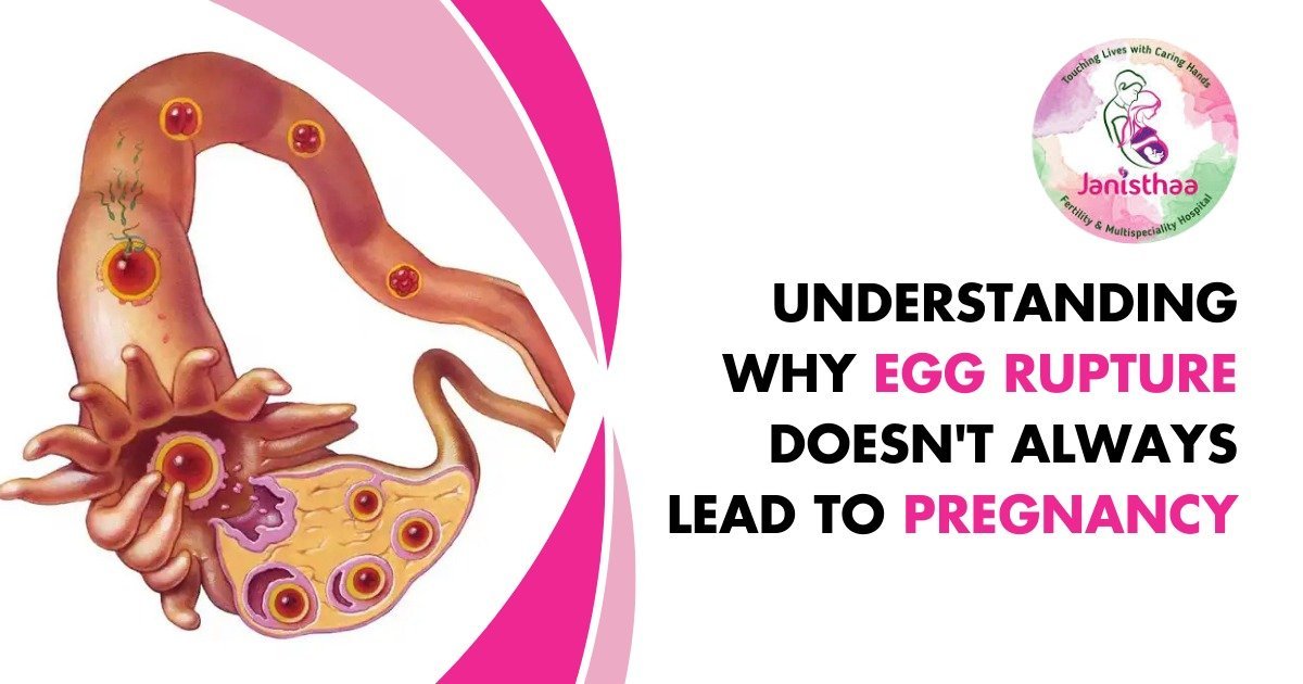
A follicular study, also called folliculometry or follicle tracking, is a series of transvaginal ultrasound (TVS) scans done at specific intervals during your menstrual cycle. The purpose is to watch your ovarian follicles — tiny fluid-filled sacs inside your ovaries, each containing a developing egg — as they grow, and to identify the precise moment one is ready to release that egg (ovulation).
Timing is everything in fertility. The egg survives for only 12–24 hours after ovulation. Sperm can live 48–72 hours. That means the fertile window is very narrow — approximately 3–5 days per cycle. Without follicular monitoring, couples and doctors are essentially guessing at timing. With it, the window is tracked in real time, every 2–3 days, until the exact date of ovulation is confirmed or predicted.
Who Needs Follicular Monitoring?
You likely need a follicular study if you are:
- Trying to conceive for 6+ months without success
- On ovulation induction medication (Letrozole / Clomiphene)
- Undergoing IUI treatment
- Preparing for or in an IVF stimulation cycle
- Diagnosed with PCOS, irregular periods, or anovulation
- Over 35 and trying to conceive
- Having cycles longer than 35 days or shorter than 21 days
What follicular monitoring tells your doctor:
- Whether you are ovulating in this cycle
- Whether the dominant follicle is growing at the right rate
- The exact predicted date of ovulation
- Whether ovulation actually occurred (follicle ruptured)
- The thickness and quality of the uterine lining
- How well ovulation-inducing medication is working
- Whether OHSS risk is developing (in IVF cycles)
Day-by-Day Scan Schedule — What Happens at Each Visit
A typical follicular study cycle involves 4–6 scans spread across approximately 10–14 days. Each scan takes 15–20 minutes and uses the transvaginal route for the clearest view of the follicles.
Establishing your starting point
The cycle begins with a baseline scan on Day 2 or Day 3 of menstruation. This assesses: the resting appearance of both ovaries, the antral follicle count (AFC — number of small follicles visible), the uterine lining (which should be thin, around 4–6 mm at this stage), and any ovarian cysts that need to be resolved before treatment begins. This scan determines whether stimulation medication can be started and what dose is appropriate.
Watching for the dominant follicle
By Day 9–10, follicles that are going to develop in this cycle become visible and measurable (typically 8–12 mm). The doctor identifies the dominant follicle — the one growing fastest that will likely ovulate — and measures all visible follicles in both ovaries. The endometrial lining thickness is also recorded. In most women, one dominant follicle begins to emerge at this point while others remain small.
The follicle is growing 1–3 mm per day
During the active growth phase, the dominant follicle grows approximately 1–3 mm per day. Scans are done every 2–3 days (or daily if the follicle is close to mature). When the follicle reaches approximately 16–18 mm, the doctor prepares for the trigger injection or advises the optimal timing for intercourse or IUI. The uterine lining should now be thickening towards 7–10 mm in a trilaminar (triple-layer) pattern.
Follicle at 18–25 mm — ready to release
Once the dominant follicle reaches 18–20 mm, it is considered mature and capable of releasing a healthy egg. At this point, the doctor may administer an hCG trigger injection (typically ovitrelle or HCG 5000–10000 IU) to trigger ovulation within 34–36 hours. The timing of intercourse, IUI, or IVF egg retrieval is calculated from this trigger point. The lining should ideally be ≥8 mm at this stage.
Confirming the egg was released
The final scan in the cycle confirms whether ovulation actually occurred. Signs of successful ovulation: the dominant follicle has collapsed (disappeared or shrunk significantly), a small amount of free fluid is visible in the pouch of Douglas (behind the uterus), and the corpus luteum (a cyst-like structure) has formed in the ovary. If the follicle is still intact at this scan, it may be an unruptured follicle (LUF) — which requires further evaluation.
Follicle Size Chart — What's Normal at Each Stage
| Cycle Day (28-day cycle) | Dominant Follicle Size | Status | Clinical Action |
|---|---|---|---|
| Day 2–3 (baseline) | 2–8 mm (antral follicles) | Resting — normal | Baseline scan, start medication if indicated |
| Day 9–10 | 8–12 mm | Early growth — normal | Confirm dominant follicle emerging, continue monitoring |
| Day 11–12 | 12–16 mm | Active growth — on track | Scan every 2 days, watch lining thickness |
| Day 13–14 | 16–18 mm | Approaching mature — near trigger zone | Plan trigger injection timing, advise timed intercourse or IUI timing |
| Day 14–16 ✅ | 18–25 mm | Mature — ready for ovulation | Trigger injection given or natural ovulation imminent. IUI / intercourse within 24–36 hours |
| Day 18+ (follicle still growing) | >25 mm — not rupturing | Unruptured — concern | Check for LUF syndrome; discuss intervention with doctor |
| No dominant follicle seen by Day 12 | <10 mm, none developing | Anovulatory cycle — abnormal | Investigate — PCOS, low AMH, medication non-response |
Follicle growth rate: In a stimulated cycle (on Letrozole or gonadotropins), the dominant follicle typically grows 1–3 mm per day. In a natural unstimulated cycle, growth may be slightly slower. Your doctor adjusts monitoring frequency based on this rate — once a follicle reaches 14–16 mm, scans are usually done every day or every other day.
Endometrial Thickness During Follicular Study — Why It Matters
Every follicular study scan also measures the uterine lining (endometrium). A thick, well-developed lining is essential for the embryo to implant successfully. The lining must be in the right condition at the time of ovulation — regardless of follicle size.
| Endometrial Thickness at Ovulation | Pattern | Interpretation |
|---|---|---|
| 8–12 mm ✅ | Trilaminar (triple-layer) | Optimal — best implantation rates |
| 7–8 mm | Trilaminar or bilayered | Acceptable — IUI or timed intercourse can proceed |
| Below 7 mm | Thin, homogeneous | Thin lining — may need investigation or oestrogen support before embryo transfer |
| Below 6 mm | Thin | Concerning — IUI or embryo transfer typically deferred; treatment to improve thickness recommended |
What causes a thin uterine lining? Poor blood flow to the uterus, low oestrogen, previous uterine surgery or D&C, Asherman’s syndrome, or certain medications. If your lining is consistently thin during follicular monitoring, Dr. Shwetha may recommend oestrogen supplementation, low-dose aspirin (to improve blood flow), or further investigation before proceeding with IUI or IVF.
Reading Your Follicular Study Report — Normal vs Abnormal
| Finding on Report | What It Means | What Happens Next |
|---|---|---|
| Dominant follicle 18–25 mm, lining 8–12 mm trilaminar, free fluid in pouch of Douglas | Ovulation has occurred — excellent cycle | Timed intercourse / IUI timing confirmed; beta hCG test in 14 days |
| Multiple follicles 15–20 mm after gonadotropin stimulation (IVF cycle) | Good ovarian response — ready for egg retrieval | Trigger injection given; egg retrieval scheduled 34–36 hours later |
| Single dominant follicle, slow growth (less than 1 mm/day) | Poor follicle development — may need medication adjustment | Increase stimulation dose or switch medication; review AMH |
| Dominant follicle reached 20mm but still present on next scan (not ruptured) | Luteinised Unruptured Follicle (LUF) | Trigger injection recommended; investigate recurring LUF |
| No dominant follicle developing by Day 14 (all <10 mm) | Anovulatory cycle — no ovulation this cycle | Investigate for PCOS, hypothyroidism, poor ovarian reserve; consider medication |
| Many small follicles (>12, each <10 mm), no dominant one, “string of pearls” appearance | PCOS pattern | Confirm PCOS diagnosis; adjust treatment; monitor carefully for OHSS risk |
| Thin lining (<7mm) at time of mature follicle | Endometrial receptivity issue | Consider oestrogen supplementation; defer IUI/embryo transfer if lining does not improve |
Role of Follicular Study in IVF and IUI
In IUI (Intrauterine Insemination)
Follicular monitoring is essential in every IUI cycle — natural or stimulated. The IUI procedure must be performed within 12–36 hours of ovulation for any chance of success. Without follicular study, the timing is guesswork. With it, the window is precisely identified. At Janisthaa IVF, every IUI cycle includes follicular monitoring as standard. Once the dominant follicle reaches 18–20 mm, the trigger injection is given and the IUI is scheduled exactly 36 hours later.
In IVF Stimulation Cycles
In IVF, the goal is different — instead of one egg, the protocol aims to stimulate multiple follicles to grow simultaneously. Follicular monitoring during an IVF stimulation cycle serves to:
- Track how many follicles are developing and their growth rate
- Adjust the stimulation dose daily based on follicle response
- Identify the optimal moment for egg retrieval — when most follicles are 17–20 mm
- Monitor for Ovarian Hyperstimulation Syndrome (OHSS) risk — if too many follicles develop (especially in PCOS patients), the retrieval may be delayed or a freeze-all strategy used
- Confirm endometrial readiness before a fresh embryo transfer
How many scans in an IVF cycle? In a stimulated IVF cycle, follicular monitoring scans are typically done every 2 days from Day 2, then daily once follicles approach 14 mm. Most IVF cycles require 5–8 monitoring scans across the stimulation period. At Janisthaa IVF, all stimulation monitoring is conducted at our three Bangalore centres and results are reviewed by Dr. Shwetha personally.
Follicular Study and PCOS — What's Different
PCOS (Polycystic Ovary Syndrome) is the most common cause of ovulatory infertility, and follicular monitoring takes on a particularly important role in PCOS management.
| Aspect | Normal Cycle | PCOS Cycle |
|---|---|---|
| Antral follicle count (baseline) | 5–15 follicles per ovary | 12+ follicles per ovary (“string of pearls”) |
| Dominant follicle development | One clear dominant follicle by Day 10–12 | Multiple small follicles competing; no clear dominant; may arrest |
| Ovulation | Predictable — Day 14–16 in a 28-day cycle | Unpredictable or absent — cycles may be 35–90 days |
| Response to medication | Usually responds well to low-dose Letrozole | May over-respond — OHSS risk; requires very close monitoring |
| Endometrial pattern | Normal trilaminar pattern by Day 14 | May be thin or irregular — particularly with long anovulatory cycles |
At Janisthaa IVF Bangalore, PCOS patients on ovulation induction are monitored especially carefully — typically with more frequent scans and lower medication doses to reduce OHSS risk. Dr. Shwetha uses individualised protocols for every PCOS patient rather than a standard dose schedule.
Follicular Study Cost in Bangalore 2026
| Type of Scan | Cost at Standalone Lab | Cost at Fertility Clinic | Notes |
|---|---|---|---|
| Single follicular scan (one visit) | ₹500 – ₹1,000 | ₹800 – ₹1,500 | Each individual scan visit |
| Complete follicular study (3 visits) | ₹1,500 – ₹2,500 | ₹2,000 – ₹4,000 | Most natural cycle monitoring |
| Complete follicular study (5–6 visits) | ₹2,500 – ₹5,000 | ₹3,000 – ₹6,000 | Stimulated cycles / PCOS cycles |
| IVF stimulation monitoring (all scans) | Varies | Included in IVF package at Janisthaa | All monitoring scans in an IVF cycle included |
Dr. Shwetha Y Baratikkae — on follicular monitoring at Janisthaa IVF
“Follicular monitoring is not just a scan — it is a real-time conversation between the patient’s body and the treatment plan. Every number we get at each visit — the follicle size, the lining thickness, the growth rate — shapes what we do next. I personally review every monitoring report before adjusting medication doses or scheduling a trigger. It is the most individualised aspect of fertility treatment, and it should never be rushed.”
20+ years experience | 10,000+ successful pregnancies | IVF Specialist, Janisthaa IVF Bangalore
Ready to start your fertility monitoring at Janisthaa IVF?
Book a consultation with Dr. Shwetha — personalised follicular monitoring across all 3 Bangalore locations
FAQs
1. What is a follicular study can?
A follicular study (folliculometry) is a series of transvaginal ultrasound scans performed every 2–3 days from Day 9 of your menstrual cycle. Each scan measures the size of ovarian follicles to predict ovulation — the exact moment an egg is released. It is the most accurate method to time natural conception, IUI, and IVF egg retrieval.
2.What is the cost of follicular study in Bangalore?
A single follicular scan in Bangalore costs ₹500–₹1,000 at diagnostic centres. A complete monitoring cycle (3–5 scans) costs ₹1,500–₹4,000 depending on the number of visits required. At Janisthaa IVF Center, follicular monitoring is included within ovulation induction and IVF treatment packages. Call +91 95911 11407 for current pricing.
3. On which day of the menstrual cycle is the follicular study done?
The follicular study begins with a baseline scan on Day 2 or 3 of your menstrual cycle. Regular monitoring scans start from Day 9 or Day 10, continuing every 2–3 days (and daily as ovulation approaches) until the follicle reaches 18–20 mm and ovulation occurs — typically between Day 12 and Day 18 in a 28-day cycle.
4.What size follicle is needed for ovulation?
A dominant follicle must reach 18–25 mm to be considered mature. At 18–20 mm, it is ready for a trigger injection (hCG) which causes ovulation within 34–36 hours. Ovulation can also occur spontaneously at this size in natural cycles. Follicles below 18 mm are not yet mature enough to release a viable egg.
5.What happens if the follicle does not rupture?
If a mature follicle fails to rupture and release the egg, it is called a Luteinised Unruptured Follicle (LUF). The follow-up scan will show the follicle still present despite the expected ovulation window having passed. Your doctor may recommend an hCG trigger injection to induce ovulation, or investigate underlying causes such as PCOS or endometriosis.
6.Is a follicular study scan painful?
A transvaginal follicular scan is generally not painful. Most women feel mild pressure or slight discomfort during the scan, which lasts approximately 10–20 minutes. No anaesthesia is needed. No dietary restrictions apply. You do not need a full bladder for a transvaginal scan — the opposite is true, an empty bladder is more comfortable.
7.How many scans are needed in a follicular study?
A typical follicular study involves 3–6 scans: Day 2–3 baseline, Day 9–10 first monitoring, Day 11–13 active growth phase scans (every 2 days), a pre-ovulation scan near Day 14–16, a post-trigger confirmation scan, and a final scan to confirm rupture. In PCOS or stimulated cycles, more frequent scans may be needed.
8.What is the difference between a follicular study and AMH test?
AMH (Anti-Müllerian Hormone) is a blood test that measures your overall ovarian reserve — how many eggs you have remaining. A follicular study is a dynamic scan-based process that watches how follicles actually grow and release eggs in a real cycle. AMH answers “how many eggs do I have left?” while follicular monitoring answers “are my eggs growing and releasing correctly this cycle?” Both tests are complementary and used together in fertility evaluations.
9.Can a follicular study detect PCOS?
Yes. In PCOS, a follicular scan shows the characteristic appearance of 12 or more small follicles (each under 10 mm) arranged around the ovary — known as the “string of pearls” sign — with no dominant follicle developing to maturity. Follicular monitoring also confirms whether a woman with PCOS is actually ovulating in a given cycle, which home ovulation kits often miss.
10. What is a good endometrial thickness during follicular study?
At the time of ovulation, an endometrial (uterine lining) thickness of 8–12 mm with a trilaminar (triple-layer) pattern is considered optimal for implantation. A lining below 7 mm is considered thin. If your lining is consistently thin during monitoring cycles, Dr. Shwetha may recommend oestrogen supplementation, low-dose aspirin, or further investigation before IUI or embryo transfer is attempted.